
Introduction
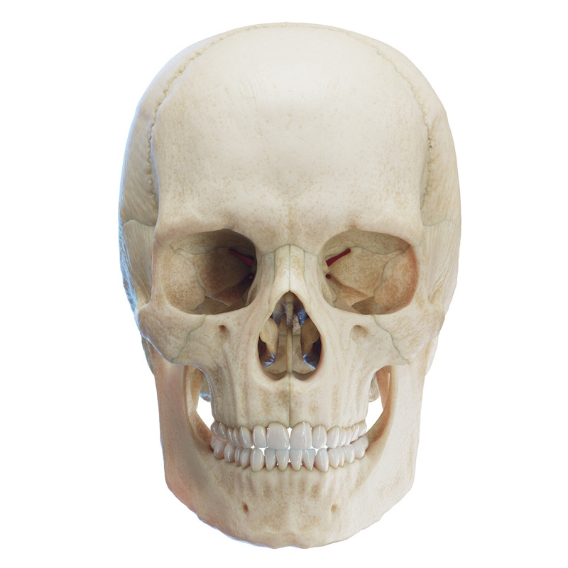
The lower jaw (mandible) is an unpaired U-shaped (as commonly described in English-language literature) or horseshoe-shaped bone. It belongs to the facial skeleton (viscerocranium) and is the largest bone in this group. The mandible is the only movable bone of the skull. It articulates with the two temporal bones (ossa temporalia) via the temporomandibular joints (TMJs).
Like the upper jaw, the lower jaw plays a critical role in facial aesthetics, forming the lower third of the face. Numerous muscles - both of mastication and of facial expression - attach to it. The mandible houses the primary and permanent dentition and plays a fundamental role in mastication (chewing), facial expression, and speech.
The mandible consists of two main parts:
- Body (corpus mandibulae): The horizontal portion of the lower jaw, which can be divided into a base (basis mandibulae) and an alveolar process/part (processus alveolaris / pars alveolaris). The body has an external and an internal surface, as well as a superior and an inferior border.
- Ascending Ramus (ramus mandibulae): The vertical portion, which forms an angle of nearly 90 degrees with the body at the angle of the mandible (angulus mandibulae / gonial angle). Depending on sex and individual anatomical variations, this angle can range between 110 and 130 degrees. It is generally larger in males.
Body of the Mandible (Corpus mandibulae)
Base of the Mandible (Basis mandibulae)
The base is the inferior, thickest portion of the mandible. In cephalometric analysis, the lowest midline point on the inferior border of the mandibular base is called the gnathion.
Features of the external surface (facies anterior/externa) of the mandibular base:
- Mental protuberance (Protuberantia mentalis): A triangular bony elevation in the midline of the body that forms the chin.
- Mental tubercle (Tuberculum mentale): A small bony prominence located on either side of the mental protuberance.
- Mental foramen (Foramen mentale): A large opening typically located in the region below the premolars. The mental nerve (nervus mentalis), which innervates the lower lip and chin on the ipsilateral side, exits through this foramen. Occasionally, more than one mental foramen may be present.
- External oblique line (Linea obliqua externa): A ridge that continues posteriorly into the temporal crest (crista temporalis). Anteriorly, it typically fades out just behind the mental foramen.
Features of the internal surface (facies posterior/interna) of the mandibular base:
- Superior and inferior mental spines (Spina mentalis superior et inferior / Genial tubercles): Two small, paired bony elevations located in the midline of the internal surface. They are directed toward the tongue and serve as the attachment sites for the genioglossus and geniohyoid muscles.
- Digastric fossa (Fossa digastrica): A paired oval depression located inferior to the mental spines on both sides of the midline. It serves as the attachment site for the anterior belly of the digastric muscle (m. digastricus).
- Mylohyoid groove (Sulcus mylohyoideus): An oblique groove extending from the posterosuperior aspect of the ascending ramus down to the anteroinferior aspect of the body.
- Mylohyoid line (Linea mylohyoidea): A ridge located superior to the mylohyoid groove. It begins just below the third mandibular molar and runs anteroinferiorly. It serves as the attachment site for the mylohyoid muscle (m. mylohyoideus). The superior pharyngeal constrictor muscle (m. constrictor pharyngis superior) attaches to the posterior portion of this line.
- Submandibular fossa (Fossa submandibularis): A paired depression located below the middle section of the mylohyoid line; it houses the submandibular gland (glandula submandibularis).
- Sublingual fossa (Fossa sublingualis): A paired depression located in the premolar region above the medial end of the mylohyoid line; it houses the sublingual gland (glandula sublingualis).
Alveolar Process (Processus alveolaris)
Located on the superior aspect of the mandibular body, it houses the teeth. The superior border of this process is called the alveolar arch (arcus alveolaris).
Features of the alveolar process:
- Dental alveoli (Alveoli dentales): Sockets, usually 16 in number per jaw, in which the teeth are anchored. Their shape, size, and number vary according to the morphology and number of the respective teeth.
- Interdental septa (Septa interalveolaria): Bony ridges separating adjacent dental alveoli.
- Interradicular septa (Septa interradicularia): Bony ridges located between the roots of a multi-rooted tooth.
- Alveolar juga (Juga alveolaria): Elevations on the external (buccal/vestibular) surface of the mandible caused by the prominence of the underlying tooth roots within their alveoli.
- Retromolar triangle (Trigonum retromolare): A triangular space formed between the third molar (wisdom tooth) and the ascending ramus.
- Retromolar fossa (Fossa retromolaris): A depression situated within the area of the retromolar triangle.
Ascending Ramus of the Mandible (Ramus mandibulae):
The ascending ramus is the paired vertical portion connected to the body, located bilaterally. It possesses two processes - the condylar process (processus condylaris) and the muscular/coronoid process (processus coronoideus) - as well as internal and external surfaces.
- Codonylar process (Processus condylaris): This is the large, rounded posterior process of the ramus. It articulates within the mandibular fossa (fossa mandibularis) and against the articular tubercle (tuberculum articulare), thereby forming the temporomandibular joint (TMJ / articulatio temporomandibularis). This process has three distinct parts.
- Head (Caput mandibulae): The uppermost rounded portion.
- Neck (Collum mandibulae): The narrow portion just below the head.
- Pterygoid fovea (Fovea pterygoidea): A depression located on the anteromedial aspect of the mandibular neck/head. The lateral pterygoid muscle (m. pterygoideus lateralis) inserts here.
- Coronoid process (Processus coronoideus): A triangular process (its name derives from its resemblance to a crown). Its anterior border is wider than its posterior border. The temporalis muscle (m. temporalis) attaches to its anterior surface and the temporal crest (crista temporalis).
- Mandibular notch (Incisura mandibulae): A deep concavity that separates the two processes of the ascending ramus.
Features of the internal surface (facies interna) of the ascending ramus:
- Mandibular foramen (Foramen mandibulae): An opening located approximately midway between the anterior and posterior borders of the ascending ramus. It leads into the mandibular canal (canalis mandibulae). It houses the inferior alveolar nerve (n. alveolaris inferior), which innervates the mandibular teeth on that side. The mental nerve (n. mentalis) branches off from it to innervate the chin, lower lip, and vestibular mucosa upon exiting the mental foramen. The inferior alveolar nerve then continues anteriorly as the incisive nerve (n. incisivus), which innervates the anterior (frontal) teeth.
- Lingula (Lingula mandibulae): A triangular bony spine that partially overlaps the opening of the mandibular foramen. The sphenomandibular ligament (ligamentum sphenomandibulare) attaches to it.
- Mylohyoid groove (Sulcus mylohyoideus): A small groove that extends anteriorly and inferiorly from the mandibular foramen. The mylohyoid nerve and artery (n. mylohyoideus, a. mylohyoidea) pass through it, associated with the mylohyoid muscle.
- Pterygoid tuberosity (Tuberositas pterygoidea): A roughened area on the internal surface of the ramus, located posteroinferior to the mylohyoid groove. The medial pterygoid muscle (m. pterygoideus medialis) inserts here.
Features of the external surface (facies externa) of the ascending ramus:
- Masseteric tuberosity (Tuberositas masseterica): A roughened bony surface on the external aspect of the ascending ramus near the angle, serving as the attachment point for the masseter muscle (m. masseter). A corresponding depression in this same region is known as the masseteric fossa (fossa masseterica).