
Introduction
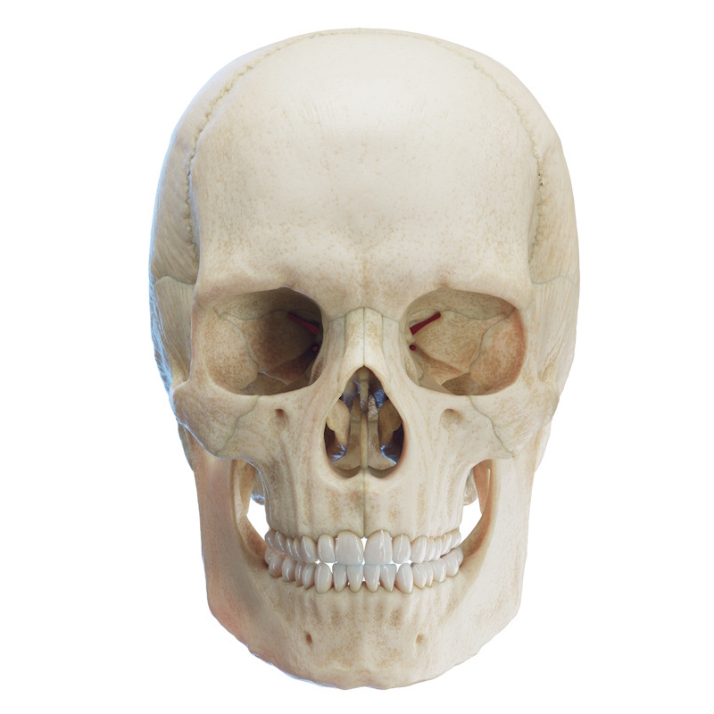
The temporal bone (os temporale) is a paired, irregularly shaped bone of the neurocranium situated on the lateral aspect of the skull inferior to the parietal bone (os parietale). Forming part of the lateral skull base (basis cranii), it is one of the most structurally complex bones in the human body. It houses the organs of hearing and balance (middle and inner ear) and is traversed by numerous canals transmitting major neurovascular structures.
The temporal bone contributes to the formation of the temporal fossa (fossa temporalis), middle cranial fossa (fossa cranii media), and posterior cranial fossa (fossa cranii posterior). It houses the three auditory ossicles: the malleus (hammer), incus (anvil), and stapes (stirrup).
Articulations and Attachments:
- Articulations (5): Parietal bone, occipital bone (os occipitale), sphenoid bone (os sphenoidale), zygomatic bone (os zygomaticum), and the mandible (mandibula) via the temporomandibular joint (TMJ).
- Muscular Attachments: Temporalis, sternocleidomastoid (SCM), digastric (posterior belly), stylohyoid, splenius capitis, longissimus capitis, stylopharyngeus, and styloglossus.
Major Parts of the Temporal Bone
The temporal bone is divided into four distinct anatomical parts:
- Squamous part (pars squamosa): A thin, flat, vertical plate forming the anterosuperior lateral wall of the skull.
- Tympanic part (pars tympanica): A small, curved bony plate situated inferolateral to the squama and anterior to the mastoid process, enclosing the external acoustic meatus.
- Mastoid part (pars mastoidea): Positioned posteroinferiorly, dominated by the easily palpable mastoid process (processus mastoideus).
- Petrous part (pars petrosa / pyramid): A dense, wedge-shaped pyramidal mass projecting anteromedially into the cranial base.
Note: Some authors group the petrous and mastoid parts together as the petromastoid part. For clarity, they are detailed separately below.
Squamous Part (Squama temporalis / Pars squamosa)
The squamous part is the largest component, forming a thin vertical plate with two primary surfaces and two borders:
Surfaces
- Temporal surface (facies temporalis): Smooth and convex lateral surface forming part of the temporal fossa; gives origin to the temporalis muscle (m. temporalis).
- Cerebral surface (facies cerebralis): Concave internal surface facing the temporal lobe of the brain, contributing to the middle cranial fossa. Marked by gyral/digital impressions (impressiones digitatae) and arterial grooves (sulci arteriosi) for the middle meningeal vessels.
Borders
- Parietal border (margo parietalis): Superior and posterior border articulating with the parietal bone. Its posterior terminus features the parietal notch (incisura parietalis), a critical surgical landmark identifying the anterior boundary of the sigmoid sinus.
- Sphenoidal border (margo sphenoidalis): Anteroinferior serrated border articulating with the greater wing of the sphenoid bone.
Anatomical landmarks of the Temporal Surface
- Zygomatic process (processus zygomaticus): Projects anteriorly to articulate with the temporal process of the zygomatic bone, completing the zygomatic arch (arcus zygomaticus).
- Suprameatal crest (crista suprameatica): A horizontal ridge continuous with the posterior root of the zygomatic process above the external acoustic meatus.
- Supramastoid crest (crista supramastoidea): Posterior continuation of the suprameatal crest running backward over the mastoid region.
- Suprameatal triangle / fossa (fovea suprameatica / Macewen's triangle): A small depression posterosuperior to the external acoustic meatus, serving as a landmark for the underlying mastoid antrum.
- Suprameatal spine (spina suprameatica / Henle's spine): A small, inconstant bony projection at the anterior margin of the suprameatal triangle.
- Mandibular fossa (fossa mandibularis): A deep depression inferior to the root of the zygomatic process that receives the condyle (caput) of the mandible to form the temporomandibular joint. Divided into an anterior articular surface (facies articularis) and a posterior non-articular part.
- Articular tubercle (tuberculum articulare): A prominent bony eminence anterior to the mandibular fossa that provides attachment for the lateral TMJ ligament.
- Articular eminence (eminentia articularis): The smooth, rounded anterior slope of the articular tubercle over which the mandibular condyle glides during jaw opening.
- Groove for middle temporal artery (sulcus a. temporalis mediae): A vertical groove extending superiorly above the external acoustic meatus.
Tympanic Part (Pars tympanica)
The smallest division, situated inferior to the squama, anterior to the mastoid process, and lateral to the petrous part. It forms the bony walls of the external acoustic meatus (meatus acusticus externus), a 2–3 cm canal closed medially by the tympanic membrane (membrana tympani).
Anatomical landmarks
- Tympanic ring (annulus tympanicus): An incomplete C-shaped bony ring that holds the periphery of the tympanic membrane.
- Tympanic sulcus (sulcus tympanicus): A narrow groove within the tympanic ring anchoring the fibrous annulus of the eardrum.
- Greater and lesser tympanic spines (spina tympanica major et minor): Bony projections at the anterosuperior and posterosuperior ends of the tympanic ring, bounding the tympanic notch (incisura tympanica / Notch of Rivinus).
- Sheath of the styloid process (vagina processus styloidei): A sharp, inferiorly extending bony margin enclosing the base of the styloid process.
- Fissures: The tympanosquamous fissure (fissura tympanosquamosa) separates the tympanic part from the squamous part; medially splits into the petrotympanic fissure (fissura petrotympanica / Glasserian fissure) (transmitting the chorda tympani nerve) and the petrosquamous fissure (fissura petrosquamosa). The Tympanomastoid fissure (fissura tympanomastoidea) separates the tympanic part from the mastoid process; transmits the auricular branch of the vagus nerve (Arnold's nerve).
Mastoid Part (Pars mastoidea)
Positioned posteroinferiorly, the mastoid part is primarily formed by the mastoid process (processus mastoideus), a conical projection pointing downward that serves as an attachment site for the sternocleidomastoid, splenius capitis, longissimus capitis, and posterior belly of the digastric muscles.
Anatomical Features
- Mastoid process (processus mastoideus): A thick, conical bony projection extending inferiorly. It is easily palpable posterior to the auricle (pinna). Its external surface is rough to serve as an attachment site for several muscles (e.g., sternocleidomastoid, splenius capitis, longissimus capitis).
- Mastoid notch (incisura mastoidea / digastric groove): A deep groove situated on the medial aspect of the mastoid process that gives origin to the posterior belly of the digastric muscle (m. digastricus).
- Groove for occipital artery (sulcus arteriae occipitalis): A shallow groove located medial to the mastoid notch through which the occipital artery (a. occipitalis) passes.
- Mastoid foramen (foramen mastoideum): An opening located near the posterior border of the mastoid portion that transmits the mastoid branch of the occipital artery (as well as a mastoid emissary vein to the sigmoid sinus).
- Mastoid antrum (antrum mastoideum): An irregularly shaped air cavity in the superior aspect of the mastoid portion. Anteriorly, it communicates with the epitympanic recess and the tympanic cavity of the middle ear. Posteroinferiorly, it communicates with the mastoid air cells (cellulae mastoideae). The roof of the mastoid antrum is formed by the tegmen mastoideum, which continues anteriorly as the tegmen tympani of the petrous part.
- Mastoid air cells (cellulae mastoideae): Numerous air-filled intercommunicating spaces within the mastoid process that vary in size, shape, and number. They open into the mastoid antrum and communicate with the middle ear, serving as air reservoirs and assisting in pressure regulation.
Petrous Part (Pars petrosa / Pyramid)
A dense, wedge-shaped pyramidal part projecting anteromedially between the sphenoid and occipital bones, separating the middle cranial fossa from the posterior cranial fossa. It houses the delicate inner ear structures and contains numerous internal canals.
Borders
- Anterior border (margo anterior): Articulates laterally with the squamous part and medially with the greater wing of the sphenoid bone.
- Superior border / Petrous crest (crista petrosa): Gives attachment to the tentorium cerebelli and features the sulcus for the superior petrosal sinus (sulcus sinus petrosi superioris).
- Posterior border (margo posterior): Articulates with the basilar and lateral parts of the occipital bone; features the jugular notch (incisura jugularis), which forms the anterolateral boundary of the jugular foramen (foramen jugulare).
Cavities & Labyrinths of the Petrous Part
Bony Tympanic Cavity (Cavitas tympani)
An air-filled middle ear space lined with mucosa, situated between the tympanic membrane and the inner ear. It houses:
- Auditory Ossicles: Malleus, incus, and stapes.
- Intratympanic Muscles: Tensor tympani (m. tensor tympani) and stapedius (m. stapedius).
- Neurovascular Structures: Chorda tympani nerve, tympanic plexus (plexus tympanicus), and mucosal mucosal folds.
Inner Ear (Auris interna)
Houses the vestibulocochlear organ (organum vestibulocochleare) responsible for hearing and balance, consisting of:
- Bony Labyrinth (labyrinthus osseus): Contains the vestibule (vestibulum), three semicircular canals (canales semicirculares), and the cochlea.
- Membranous Labyrinth (labyrinthus membranaceus): Suspended within the bony labyrinth and filled with endolymph. Contains the saccule, utricle, semicircular ducts, and cochlear duct (ductus cochlearis). The perilymphatic space (spatium perilymphaticum) is the fluid-filled space containing perilymph situated between the bony and membranous labyrinths.
Apex of the Petrous Part (Apex partis petrosae)
Points anteromedially to form the posterior border of the foramen lacerum (cartilage-filled in life).
- Carotid canal (canalis carotidis): A passageway transmitting the internal carotid artery (a. carotis interna) alongside its sympathetic and venous plexuses.
- Internal orifice of the carotid canal (apertura interna canalis carotidis): The exit opening situated at the apex of the petrous apex on the internal skull base, through which the internal carotid artery enters the cranial cavity.
- Carotidotympanic canaliculi (canaliculi carotidotympanici): Small passages connecting the carotid canal directly to the tympanic cavity, transmitting the carotidotympanic nerves and vessels.
- Musculotubal canal (canalis musculotubarius): A double-barreled channel divided into two semicanals by a thin bony partition. Its external orifice lies on the inferior surface of the petrous part anterior to the external carotid opening, while its internal orifice leads into the tympanic cavity.
- Semicanal for tensor tympani (semicanalis m. tensoris tympani): The smaller, superior compartment of the musculotubal canal, housing the tensor tympani muscle (m. tensor tympani).
- Semicanal for auditory tube (semicanalis tubae auditivae): The larger, inferior compartment of the musculotubal canal, forming the osseous portion of the Eustachian (auditory) tube.
- Septum of the musculotubal canal (septum canalis musculotubarii): The thin, horizontal bony shelf separating the musculotubal canal into its superior and inferior semicanals.
Anterior Surface of the Petrous Part (Facies anterior partis petrosae)
The anterior surface of the petrous part forms part of the floor of the middle cranial fossa (fossa cranii media).
- Tegmen tympani: A smooth, thin bony plate situated anterolaterally on the anterior surface that forms the roof of the tympanic cavity (middle ear) and mastoid antrum, separating the tympanic cavity from the cranial cavity. It is located anterior and lateral to the arcuate eminence.
- Arcuate eminence (eminentia arcuata): A rounded bony elevation near the center of the anterior surface produced by the underlying anterior (superior) semicircular canal of the inner ear.
- Trigeminal impression (impressio trigeminalis): A shallow depression situated medially near the petrous apex that lodges the trigeminal (Gasserian) ganglion (ganglion trigeminale).
- Hiatus for greater petrosal nerve (hiatus canalis nervi petrosi majoris): A small, oblique opening located lateral to the trigeminal impression that transmits the greater petrosal nerve (nervus petrosus major) and connects directly to the facial canal.
- Groove for greater petrosal nerve (sulcus nervi petrosi majoris): A shallow, narrow groove extending anteriorly from the hiatus for the greater petrosal nerve.
- Hiatus for lesser petrosal nerve (hiatus canalis nervi petrosi minoris): A small, oblique opening situated lateral and anterior to the hiatus for the greater petrosal nerve that transmits the lesser petrosal nerve (nervus petrosus minor).
- Groove for lesser petrosal nerve (sulcus nervi petrosi minoris): A shallow, narrow groove extending from the lesser petrosal hiatus. Running parallel to each other, the groove for the lesser petrosal nerve lies anterior/anterolateral to the groove for the greater petrosal nerve.
Posterior Surface of the Petrous Part (Facies posterior partis petrosae)
The posterior surface forms the anterior boundary of the posterior cranial fossa (fossa cranii posterior).
- Internal acoustic opening (porus acusticus internus): A large, rounded orifice located near the center of the posterior surface that leads into the internal acoustic meatus (meatus acusticus internus).
- Internal acoustic meatus (meatus acusticus internus): A short, narrow canal (approximately 1 cm in length) transmitting the facial nerve (CN VII / n. facialis), vestibulocochlear nerve (CN VIII / n. vestibulocochlearis), nervus intermedius, and labyrinthine (internal auditory) artery (a. labyrinthi).
- Subarcuate fossa (fossa subarcuata): A slight, irregularly shaped depression situated posterosuperior to the internal acoustic opening (more prominent in newborns).
- Aperture of the vestibular canaliculus (apertura canaliculi vestibuli): A small, slit-like opening situated inferior to the petrous crest and lateral to the internal acoustic meatus; leads into the vestibular canaliculus to transmit the endolymphatic duct and sac.
- Vestibular canaliculus (canaliculus vestibuli): A narrow canal extending from the inner ear's vestibule to the posterior surface of the petrous part.
Inferior Surface of the Petrous Part (Facies inferior partis petrosae)
The inferior surface is rough and irregular, contributing to the external cranial base (basis cranii externa).
- Styloid process (processus styloideus): A slender, pointed, fragile projection extending anteroinferiorly from the base of the temporal bone. Varying in length among individuals, it gives attachment to the stylohyoid muscle (m. stylohyoideus), styloglossus, stylopharyngeus, and the stylomandibular ligament (lig. stylomandibulare).
- Stylomastoid foramen (foramen stylomastoideum): A prominent opening located immediately posterior to the base of the styloid process. It represents the external terminus of the facial canal (canalis facialis), transmitting the motor trunk of the facial nerve (CN VII) and the stylomastoid artery (a. stylomastoidea).
- Facial canal (canalis facialis / fallopian canal): A Z-shaped osseous canal traversing the temporal bone that houses the facial nerve (CN VII). It begins at the internal acoustic meatus and exits at the stylomastoid foramen. From the meatus, it courses anteriorly to the hiatus for the greater petrosal nerve, makes a 90-degree sharp bend at the geniculum (geniculum canalis facialis), runs posterolaterally parallel to the petrous axis past the arcuate eminence, bends 90 degrees downward, and terminates at the stylomastoid foramen.
- Geniculum of the facial canal (geniculum canalis facialis): The sharp, 90-degree knee-like bend of the facial canal accommodating the geniculate ganglion.
- Canaliculus for chorda tympani (canaliculus chordae tympani): A tiny branch canal arising from the descending segment of the facial canal above the stylomastoid foramen, transmitting the chorda tympani nerve into the tympanic cavity.
- Jugular fossa (fossa jugularis): A deep, oval depression that forms the anterolateral wall of the jugular foramen, lodging the superior bulb of the internal jugular vein (v. jugularis interna).
- Aperture of the cochlear canaliculus (apertura canaliculi cochleae): A small opening situated on the ridge adjacent to the jugular notch and jugular fossa, leading into the cochlea.
- Cochlear canaliculus (canaliculus cochleae): A narrow duct transmitting the perilymphatic duct from the cochlea to the subarachnoid space.
- Mastoid canaliculus (canaliculus mastoideus): A tiny channel located in the lateral wall of the jugular fossa transmitting the auricular branch of the vagus nerve (CN X / Arnold's nerve).
- External orifice of the carotid canal (apertura externa canalis carotidis): The inferior entrance of the carotid canal, situated anterior to the jugular fossa.
- Petrosal fossa (fossula petrosa): A small, shallow depression situated on the bony ridge separating the external carotid orifice from the jugular fossa.
- Tympanic canaliculus (canaliculus tympanicus / Jacobson's canal): A tiny opening situated within the petrosal fossa between the carotid orifice and jugular fossa. It transmits the tympanic nerve (a branch of the glossopharyngeal nerve / CN IX) and the inferior tympanic artery (a. tympanica inferior).
- Intrajugular process of the temporal bone (processus intrajugularis ossis temporalis): A small bony spine extending into the jugular notch that may articulate with the corresponding intrajugular process of the occipital bone (processus intrajugularis ossis occipitalis) to divide the jugular foramen into neural (anteromedial) and venous (posterolateral) compartments.