
Introduction
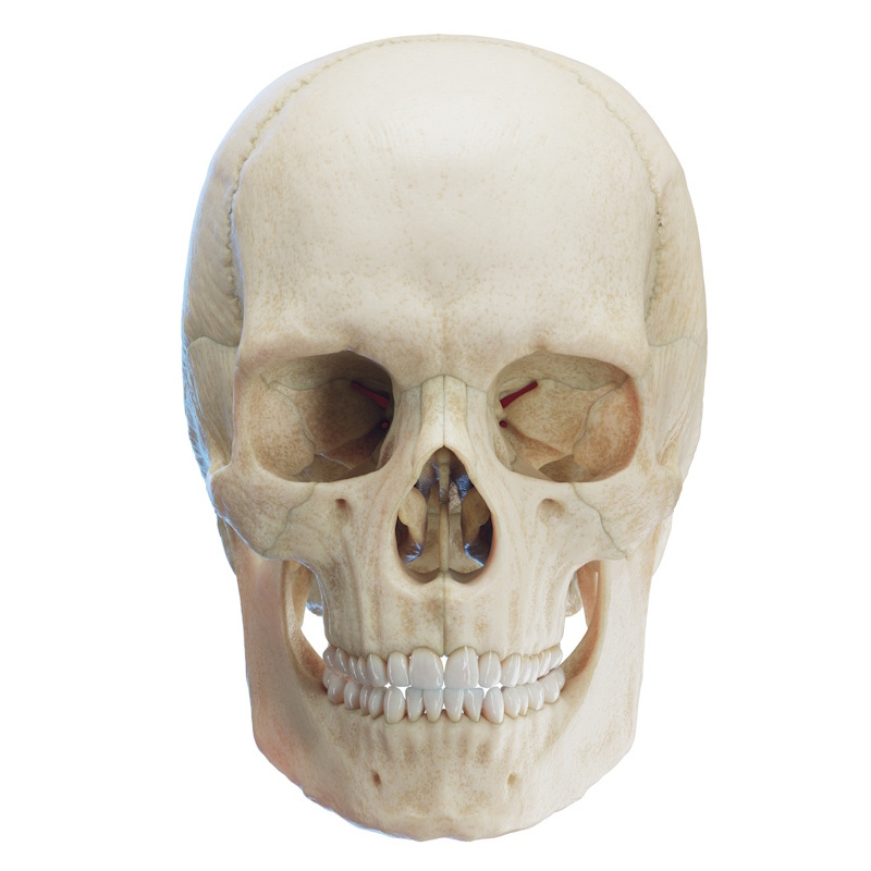
The orbits are paired bony cavities located on both sides of the root of the nose. They are situated below the anterior cranial fossa and anterior to the middle cranial fossa. Each orbit contains the eyeball, optic nerve, extraocular muscles, lacrimal apparatus, orbital fat, fascia, nerves, and vessels that supply all these structures.
Bony Orbit
Bones Forming the Orbit
The orbit is composed of seven bones that give it a pyramidal shape. The wide base faces forward towards the face, while its apex points toward the skull:
- Maxilla (upper jaw)
- Zygomatic bone (os zygomaticum)
- Frontal bone (os frontale)
- Ethmoid bone (os ethmoidale)
- Lacrimal bone (os lacrimale)
- Sphenoid bone (os sphenoidale)
- Palatine bone (os palatinum)
Bones Forming the Orbital Margins
The orbit is bounded by four margins—superior, inferior, medial, and lateral:
- Superior / Supraorbital margin (margo supraorbitalis): Formed by the orbital part of the frontal bone.
- Inferior / Infraorbital margin (margo infraorbitalis): Formed by the zygomatic process of the maxilla and the orbital process of the zygomatic bone.
- Lateral orbital margin (margo lateralis): Formed by the frontal process of the zygomatic bone.
- Medial orbital margin (margo medialis): Formed by the frontal process of the maxilla, the lacrimal bone, and the orbital part of the frontal bone.
Bones Forming the Orbital Walls
The orbit consists of four walls—superior, inferior, medial, and lateral:
- Superior wall / Roof (paries superior): Formed by the orbital surface of the frontal bone and the lesser wing of the sphenoid bone.
- Inferior wall / Floor (paries inferior): Formed by the orbital surface of the body of the maxilla, the orbital surface of the zygomatic bone, and the orbital process of the palatine bone.
- Medial wall (paries medialis): Formed by the frontal process of the maxilla, the lacrimal bone, the orbital plate of the ethmoid bone, and the body of the sphenoid bone.
- Lateral wall (paries lateralis): Formed by the orbital surface of the greater wing of the sphenoid bone and the orbital surface of the zygomatic bone.
Orbital Muscles
There are seven pairs of muscles, which include the six extraocular (oculomotor) muscles (four recti and two obliques) and the levator muscle of the upper eyelid. The rectus muscles share a common origin—they arise from a fibrous tendinous ring known as the Annulus of Zinn. This ring is located behind the eyeball at the apex of the orbital pyramid, anterior to the optic canal. These muscles travel forward, attach to various points on the sclera of the eye, and move it vertically and horizontally. The oblique muscles have an angular course and do not originate from the Annulus of Zinn, but rather from the periosteum of the regional bones; they move the eye in a sagittal direction. The extraocular musculature is unique, composed of fast fibers that are highly resistant to fatigue, and are considered the most active muscles in the human body. They continue to move even during sleep. They are also much more densely innervated than other skeletal muscles—the ratio of nerve fibers to muscle fibers is 1:3 to 1:5, compared to over 1:50 in other muscles.
Rectus Muscles
- Superior rectus muscle (m. rectus superior)
- Inferior rectus muscle (m. rectus inferior)
- Medial rectus muscle (m. rectus medialis)
- Lateral rectus muscle (m. rectus lateralis)
Oblique Muscles
- Superior oblique muscle (m. obliquus superior)
- Inferior oblique muscle (m. obliquus inferior)
Muscle Elevating the Upper Eyelid
- Levator palpebrae superioris (m. levator palpebrae superioris)
Nerves and Ganglia in the Orbit
- Optic nerve (n. opticus, Cranial Nerve II)
- Ophthalmic nerve (n. ophthalmicus, first branch of the trigeminal nerve, Cranial Nerve V1)
- Frontal nerve (n. frontalis, arises from the ophthalmic nerve)
- Supraorbital nerve (n. supraorbitalis, arises from the ophthalmic nerve)
- Supratrochlear nerve (n. supratrochlearis, arises from the ophthalmic nerve)
- Lacrimal nerve (n. lacrimalis, arises from the ophthalmic nerve)
- Nasociliary nerve (n. nasociliaris, arises from the ophthalmic nerve)
- Posterior ethmoidal nerve (n. ethmoidalis posterior, branch of the nasociliary nerve)
- Anterior ethmoidal nerve (n. ethmoidalis anterior, branch of the nasociliary nerve)
- Infratrochlear nerve (n. infratrochlearis, branch of the nasociliary nerve)
- Long ciliary nerve / nerves (n. nasociliaris longus, nn. nasociliares longi)
- Oculomotor nerve (n. oculomotorius, Cranial Nerve III)
- Ciliary ganglion (ganglion ciliare)
- Trochlear nerve (n. trochlearis, Cranial Nerve IV)
- Abducens nerve (n. abducens, Cranial Nerve VI)
Blood Vessels
Arterial Vessels
- Ophthalmic artery and its branches (a. ophthalmica)
- Infraorbital artery (a. infraorbitalis)
Venous System
The veins in the orbit do not have a valvular apparatus (valves), which facilitates the easy transmission of infections from the orbit to brain structures.
- Superior ophthalmic vein (v. ophthalmica superior)
- Inferior ophthalmic vein (v. ophthalmica inferior)
Lacrimal Apparatus
The lacrimal apparatus is involved in the production, distribution, and drainage of fluid from the surface of the eyeball (tears). It consists of a gland with excretory ducts, lacrimal canaliculi, the lacrimal sac, and the nasolacrimal duct. The lacrimal gland (glandula lacrimalis) is located in the superior and lateral part of the orbit and is divided into two parts by the levator palpebrae superioris muscle—an orbital part and a palpebral (smaller) part. The gland secretes its fluid into the superior conjunctival fornix. The flow of the fluid is continuous, moving from lateral to medial, and is spread across the eye by the act of blinking. The lacrimal secretion collects medially in the lacrimal lake (lacus lacrimalis). It then drains through the lacrimal canaliculi (superior and inferior, corresponding to the eyelids), passing into the superior and inferior lacrimal puncta (punctum lacrimale), and from there into the canaliculi lacrimales. These join the lacrimal sac. The lacrimal sac is located between the anterior lacrimal crest and posterior lacrimal crest, behind the medial palpebral ligament, and anterior to the lacrimal part of the orbicularis oculi muscle. During the movement of the latter, the lacrimal sac expands and evacuates tears from the conjunctiva.
Parts of the Lacrimal Apparatus
- Lacrimal gland and its excretory ducts (glandula lacrimalis et ductuli excretorii glandulae lacrimalis)
- Lacrimal canaliculi (canaliculi lacrimales)
- Lacrimal sac (sacculus lacrimalis)
- Nasolacrimal duct (ductus nasolacrimalis)
Orbital Fissures
Superior Orbital Fissure (Fissura orbitalis superior)
The superior orbital fissure is a slit-like opening in the posterior wall of the orbit, formed entirely by parts of the sphenoid bone. The superior border is the lesser wing, the inferior and lateral border is the greater wing, and the medial border is the body. The structures passing through it are anatomically and functionally divided into three parts—lateral, middle, and medial. This division is determined by their position relative to the common tendinous ring (Annulus of Zinn), which surrounds the optic canal and the central part of the fissure. It communicates with the middle cranial fossa.
- Lateral Part (lateral to the Annulus of Zinn):
- Trochlear nerve (n. trochlearis, CN IV)
- Frontal nerve (n. frontalis)
- Lacrimal nerve (n. lacrimalis)
- Superior ophthalmic vein (v. ophthalmica superior)
- Recurrent meningeal artery (a. meningea recurrens, branch of the lacrimal or middle meningeal artery)
- Middle Part (passing through/inside the Annulus of Zinn):
- Superior division of the oculomotor nerve (CN III) – provides motor innervation to the superior rectus muscle and the levator palpebrae superioris.
- Inferior division of the oculomotor nerve (CN III) – provides motor innervation to the medial rectus, inferior rectus, and inferior oblique muscles, as well as parasympathetic fibers to the ciliary ganglion.
- Nasociliary nerve (n. nasociliaris) – provides sensory innervation to the cornea, ciliary body, and ethmoidal sinuses.
- Abducens nerve (n. abducens, CN VI) – provides motor innervation to the lateral rectus muscle.
- Sympathetic fibers – directed to the ciliary ganglion.
- Medial Part (below the Annulus of Zinn):
- Inferior ophthalmic vein (v. ophthalmica inferior) – highly variable. It can pass through or below the Annulus of Zinn. It drains into the cavernous sinus or the pterygoid venous plexus.
Inferior Orbital Fissure (Fissura orbitalis inferior)
Unlike the superior fissure, the borders of the inferior orbital fissure are formed by several bones. The anteromedial border is formed by the maxilla and palatine bone. The posterolateral border is formed by the sphenoid bone, and the anterolateral border by the zygomatic bone. It communicates with the pterygopalatine fossa (fossa pterygopalatina) and the infratemporal fossa (fossa infratemporalis).
Structures passing through the fissure
- Infraorbital nerve (n. infraorbitalis, part of the maxillary nerve, CN V2) – passes through the corresponding groove and canal, and exits through the corresponding foramen on the anterior surface of the maxilla. Provides sensation to the lower eyelid, cheek, upper lip, and some of the upper teeth.
- Zygomatic nerve (n. zygomaticus, part of the maxillary nerve, CN V2. Divides into the n. zygomaticofacialis and n. zygomaticotemporalis) – innervates the skin of the cheek and temple, and carries parasympathetic fibers to the lacrimal nerve.
- Orbital branches of the pterygopalatine ganglion (rami orbitales) – these are small autonomic and sensory fibers innervating the periosteum of the orbit (periorbita) and Müller's muscle.
- Infraorbital artery (a. infraorbitalis) – arises from the maxillary artery (a. maxillaris) and shares a common pathway with the infraorbital nerve.
- Infraorbital vein (v. infraorbitalis) – drains blood from the floor of the orbit to the pterygoid venous plexus.
- Venous branch connecting the inferior ophthalmic vein to the pterygoid plexus – a variable alternative pathway for venous drainage.
Orbital Apex
The orbital apex (apex orbitalis) is the narrowest, most posterior zone of the orbit, where the four bony orbital walls converge. It serves as the main neurovascular junction between the intracranial cavity and the orbit.
Main Structures
Located in the anatomical region of the orbital apex (some detailed above):
- Optic canal (canalis opticus) – located in the lesser wing of the sphenoid bone, medial to the orbital apex. The optic nerve (n. opticus, CN II) and the ophthalmic artery (a. ophthalmica) pass through it.
- Superior orbital fissure (fissura orbitalis superior) – located lateral and inferior to the optic canal.
- Common tendinous ring (Annulus of Zinn / annulus tendineus communis) – the fibrous ring that encompasses the optic canal and the central part of the superior orbital fissure.