
Introduction
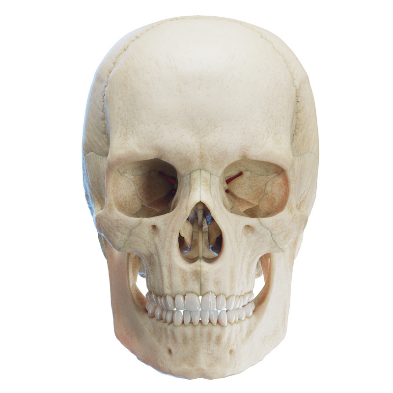
The frontal bone (os frontale) is an unpaired, bowl-shaped bone of the neurocranium. It is one of the largest and strongest bones of the skull. Its name derives from the Latin frons, meaning "forehead." Its primary function is to protect the frontal lobes of the brain. The frontal bone also plays a major structural and aesthetic role in forming the contours of the upper face, as well as contributing to the bony orbits, nasal cavity, and the floor of the anterior cranial fossa. It houses the paired frontal paranasal sinuses.
Articulations and Development:
The frontal bone articulates with 12 bones:
- Nasal bones (ossa nasalia): via the frontonasal sutures (suturae frontonasales)
- Lacrimal bones (ossa lacrimalia): via the frontolacrimal sutures (suturae frontolacrimales)
- Maxilla: via the frontomaxillary sutures (suturae frontomaxillares)
- Ethmoid bone (os ethmoidale): via the frontoethmoidal suture (sutura frontoethmoidalis)
- Sphenoid bone (os sphenoidale): via the sphenofrontal suture (sutura sphenofrontalis)
- Zygomatic bones (ossa zygomatica): via the frontozygomatic sutures (suturae frontozygomaticae)
- Parietal bones (ossa parietalia): via the coronal suture (sutura coronalis)
At birth, the frontal bone consists of two symmetrical halves separated by the frontal / metopic suture (sutura metopica). Fused usually by the second year of life, the midline suture becomes obliterated. In a small percentage of individuals, a remnant of this suture persists (most commonly in the area of the glabella) as a persistent metopic suture.
Parts of the Frontal Bone
The frontal bone consists of three main divisions:
- Squamous part (pars squamosa): The large vertical plate forming the forehead.
- Orbital part (pars orbitalis): Paired horizontal plates forming the roof of the orbits and floor of the anterior cranial fossa.
- Nasal part (pars nasalis): The small central projection extending inferiorly between the orbital plates.
Squamous Part (Pars squamosa)
The squamous part is the largest division, featuring a convex external surface (facies externa) and a concave internal cerebral surface (facies interna).
External Surface (Facies externa)
- Supraorbital margin (margo supraorbitalis): Paired curved upper boundaries of the orbits, forming the junction between the squamous and orbital parts.
- Frontal notch / foramen (incisura frontalis / foramen frontale): A small notch or foramen situated on the medial aspect of the supraorbital margin (also termed the supratrochlear notch/foramen); transmits the supratrochlear vessels and nerve.
- Supraorbital notch / foramen (incisura supraorbitalis / foramen supraorbitale): Located lateral to the frontal notch along the supraorbital margin; transmits the supraorbital vessels and nerve.
- Superciliary arch (arcus superciliaris): Paired rounded ridges located immediately superior to the supraorbital margins beneath the eyebrows; typically more prominent in males.
- Glabella: A smooth midline elevation situated between the two superciliary arches directly above the nasal bones.
- Frontal eminence / tuber (tuber frontale / eminentia frontalis): Paired rounded elevations located a few centimeters superior to the superciliary arches, marking the initial ossification centers of the bone.
- Zygomatic process (processus zygomaticus): A strong lateral projection forming the anterolateral corner of the squama; articulates with the frontal process of the zygomatic bone.
- Temporal line (linea temporalis): A ridge extending posterosuperiorly from the zygomatic process across the squama, continuous with the temporal line of the parietal bone.
- Temporal surface (facies temporalis): A smooth surface inferior to the temporal line that forms part of the temporal fossa.
- Parietal border (margo parietalis): Serrated superior margin articulating with the parietal bones at the coronal suture.
Internal Surface (Facies interna)
- Frontal crest (crista frontalis): A median vertical ridge providing attachment for the falx cerebri.
- Foramen caecum: A small, variable notch or opening at the inferior base of the frontal crest transmitting an emissary vein from the nasal cavity to the superior sagittal sinus.
- Groove for superior sagittal sinus (sulcus sinus sagittalis superioris): A vertical midline groove running superiorly from the frontal crest, lodging the superior sagittal sinus.
- Meningeal grooves (sulci meningeales): Narrow impressions accommodating the anterior branches of the middle meningeal vessels.
- Granular foveolae (foveolae granulares): Small depressions along the sagittal groove near the coronal suture that lodge arachnoid granulations (Pacchionian bodies).
- Gyral / Cerebral impressions (impressiones cerebri / digitatae): Concavities on either side of the midline corresponding to the gyri of the frontal lobes.
Orbital Part (Pars orbitalis)
The orbital part consists of paired, thin, triangular horizontal plates separated by a deep gap—the ethmoidal notch (incisura ethmoidalis) - which receives the cribriform plate of the ethmoid bone. It forms the roof of the orbit and the floor of the anterior cranial fossa.
- Orbital plate (lamina orbitalis): The triangular bony sheet constituting the orbital roof.
- Lacrimal fossa (fossa lacrimalis): A shallow depression situated anterolaterally on the orbital surface near the zygomatic process, lodging the lacrimal gland.
- Trochlear fovea (fovea trochlearis): A small depression located anteromedially on the orbital surface, serving as the attachment site for the cartilaginous pulley (trochlea) of the superior oblique muscle.
- Trochlear spine (spina trochlearis): An occasional small bony projection adjacent to the trochlear fovea.
- Sphenoidal border (margo sphenoidalis): Posterior margin that articulates with the lesser and greater wings of the sphenoid bone.
Nasal Part (Pars nasalis)
The nasal part is the smallest division, extending inferomedially between the orbital plates anterior to the ethmoidal notch.
- Nasion: The midline intersection point of the frontonasal suture, marking the junction of the frontal and nasal bones.
- Nasal spine (spina nasalis): A sharp process projecting downward and forward from the center of the nasal part, articulating with the nasal bones and the perpendicular plate of the ethmoid bone to support the nasal bridge.
- Nasal border (margo nasalis): The serrated inferior margin articulating with the nasal bones and the frontal processes of the maxillae.
Frontal Sinuses (Sinus frontales)
The frontal sinuses are paired, air-filled paranasal cavities located deep to the superciliary arches within the frontal squama.
- Varying widely in size and shape, they are typically pyramidal and separated by a thin bony partition, the inter-frontal septum (septum intersinuale frontale).
- The floor of each sinus forms part of the roof of the orbit and nasal cavity.
- They open into the middle nasal meatus (via the frontonasal duct / ethmoidal infundibulum).
- The frontal sinuses are the last paranasal sinuses to develop, being rudimentary or absent at birth and expanding significantly throughout childhood and adolescence.