
Introduction
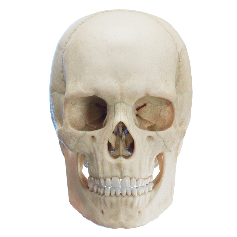
The zygomatic bone (os zygomaticum, also commonly referred to as the zygoma, cheekbone, or malar bone) is a paired, irregularly shaped bone of the viscerocranium. It forms the prominent facial contours of the cheeks, serving both aesthetic and protective structural functions. The zygomatic bone contributes to the formation of the lateral wall and floor of the orbit, the temporal fossa (fossa temporalis), and the zygomatic arch (arcus zygomaticus). It also provides an anchor site for the masseter muscle (m. masseter).
The zygomatic bone articulates with four cranial and facial bones: the maxilla, the temporal bone (os temporale), the sphenoid bone (os sphenoidale), and the frontal bone (os frontale). Anatomically, it features three processes, three surfaces, and five borders.
Processes of the Zygomatic Bone (Processi)
- Temporal process (processus temporalis): Extends posteriorly to articulate with the zygomatic process of the temporal bone, together forming the zygomatic arch (arcus zygomaticus).
- Frontal process (processus frontalis): Projects vertically superiorly to articulate with the zygomatic process of the frontal bone and the greater wing (ala major) of the sphenoid bone. It separates the orbit from the temporal fossa and forms the superior portion of the lateral orbital margin. On its orbital surface lies the orbital tubercle (protuberantia orbitalis / Whitnall's tubercle), which serves as the attachment point for the lateral palpebral ligament (ligamentum palpebrale laterale). A small backward-projecting elevation on its posterior border is known as the marginal tubercle (tuberculum marginale).
- Maxillary process (processus maxillaris): Projects anteroinferiorly to articulate with the maxilla, contributing to the lateral portion of the infraorbital margin (margo infraorbitalis). (Note: Some anatomical classifications view this area simply as the maxillary border rather than a distinct process).
Surfaces
- Orbital surface (facies orbitalis): Smooth and concave, it forms the anterolateral wall and part of the floor of the orbit. It contains the zygomatico-orbital foramen (foramen zygomaticoorbitale), which leads into a canal that bifurcates to transmit the zygomaticofacial nerve (nervus zygomaticofacialis) and zygomaticotemporal nerve (nervus zygomaticotemporalis).
- Lateral / Facial surface (facies lateralis): Convex and smooth, forming the prominent contour of the cheek. It features the zygomaticofacial foramen (foramen zygomaticofaciale), through which the zygomaticofacial nerve and vessels exit onto the face.
- Temporal surface (facies temporalis): Faces posteriorly and medially, forming part of the anterior boundary of the temporal and infratemporal fossae (fossa temporalis et fossa infratemporalis). It contains the zygomaticotemporal foramen (foramen zygomaticotemporale), transmitting the zygomaticotemporal nerve.
Borders (Margins)
- Anterosuperior (Orbital) border (margo anterosuperior): Smooth and concave; separates the orbital surface from the lateral surface and forms the inferolateral portion of the orbital rim.
- Anteroinferior (Maxillary) border (margo anteroinferior): Serrated; articulates directly with the maxilla.
- Posteroinferior border (margo posteroinferior): Thick and rough; gives attachment to the origin of the masseter muscle (m. masseter).
- Posterosuperior (Temporal) border (margo posterosuperior): Continuous with the upper border of the zygomatic arch and the posterior margin of the frontal process; gives attachment to the temporal fascia.
- Posteromedial border (margo posteromedialis): Serrated border that articulates with the greater wing of the sphenoid bone superiorly and the orbital surface of the maxilla inferiorly. It forms the lateral boundary of the inferior orbital fissure (fissura orbitalis inferior).