
Pathogenesis
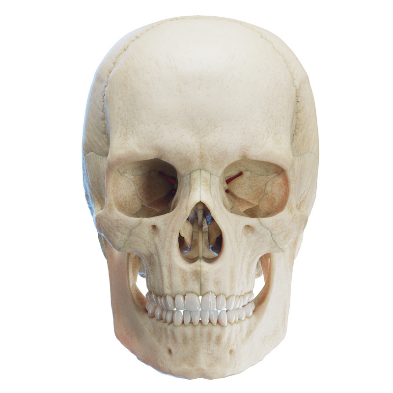
Bisphosphonates and monoclonal antibodies inhibit bone remodeling, disrupting normal bone turnover and resorption. Due to masticatory forces, jawbone metabolism is approximately ten times faster than in other skeletal sites, making the jaws pathognomonic target sites. Antiangiogenic agents and tyrosine kinase inhibitors act via distinct vascular mechanism-based pathways; temporary drug holidays often lead to rapid clinical improvement or resolution. However, combining targeted therapies with antiresorptive agents can result in an aggressive disease course.
Diagnosis
Diagnosis relies on clinical evaluation and patient symptoms, most commonly presenting as a purulent fistula or exposed bone.
Clinical Course and Severity
Patients can live with MRONJ for years. While it significantly impacts quality of life, severe abscesses or phlegmons are uncommon; the condition typically follows a chronic course.
Diagnostic Imaging
Panoramic radiograms (OPGs) are frequently performed, but clinical experience indicates they lack sufficient diagnostic sensitivity. Computed tomography (CT) or Cone-Beam Computed Tomography (CBCT) is required. CBCT can often detect pre-necrotic risk zones—such as reactive osteosclerosis—before clinical bone exposure occurs.
Role of PET Scans and Scintigraphy
Nuclear imaging modalities can detect incipient, subclinical osteonecrosis before physical manifestation, offering unmatched early diagnostic value. However, a key limitation is that affected jaw sites continue to demonstrate radiotracer uptake ("light up") even after clinical healing. Because radiologists do not routinely evaluate the jaws on general PET scans, patients with suspected necrosis should explicitly request targeted maxillofacial interpretation.
Assessing Severity
Maxillary osteonecrosis generally follows a milder course than mandibular involvement; the maxilla can occasionally self-limit and heal spontaneously, whereas the mandible rarely does. Lower lip paresthesia on the affected side of the mandible is a major warning sign, indicating either an aggressive phenotype or advanced disease stage.
Potential Complications
Infection, chronic pain, purulent drainage, halitosis/unpleasant taste, oroantral or oronasal communication (causing fluid/food regurgitation through the nose), and pathological mandibular fractures.
Treatment Modalities
- Conservative Management: Involves analgesics, systemic antibiotics, and antiseptic mouth rinses. There are no definitive standardized guidelines regarding optimal rinse formulations. Conservative protocols often rely on trial and error—adjusting medications until symptoms stabilize. Symptoms can be difficult to control, and conservative measures very rarely achieve full cure.
- Surgical Management: Aims to resect necrotic bone, relieve symptoms, or achieve disease resolution. Surgical success rates approach 100% in osteoporosis patients. In oncology patients, success rates are lower (60–70%), even with early diagnosis. Clinical experience shows that surgical intervention within the first 3–6 months yields favorable outcomes. Brief treatment delays required for drug holidays do not adversely affect surgical success. Recurrences are common, sometimes requiring repeated interventions; however, long-term remission (4+ years) is attainable. In rare instances, surgery can trigger more aggressive necrosis or pathological fracture.